Methods and Materials
Nine healthy and sub-elite female runners completed five minutes of running at 2.5 m/s. Five consecutive gait cycles were analyzed.
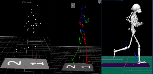
A tandem-belt instrumented treadmill (AMTI, Watertown, MA) collecting at 1000 Hz was used to collect kinetic data. A 12 camera (Vicon, United Kingdom) motion analysis system collecting at 100 Hz was used to capture locations of retroreflective markers placed over specific anatomical locations (figure 1). A three dimensional skeletal model was generated from the kinematic and kinetic data collected for each participant.
This model allowed for calculations of GRFs (figure 3) on the body and the motion of the participant’s ankle, knee, and hip joints (Figure 4). Choose this theme to customize and add content to this page.